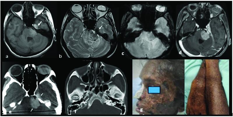
Fig. 1.

( a, b ) Axial T1WI and T2WI show T1 hyperintense and T2 hypointense lesions in left cerebellopontine angle extending to Meckel's cave ( white arrow ) suggestive of melanotic component in the lesion. Incidental finding: Deformed left globe is noted with hyperdense content on CT, mild T1 hyperintensity, and T2 hypointensity in the posterior chamber suggestive of incidental chronic vitreous hemorrhage. ( c ) SWI shows no evidence of blooming within the lesion. ( d ) Axial T1 postcontrast shows homogenous enhancement. ( e ) Axial NCCT brain shows hyperdense mass ( white arrow ). ( f ) Axial NCCT brain bone window shows smooth widening of left foramen ovale ( black arrow ). ( g ) Diffuse lentigines. CT, computed tomography; NCCT, noncontrast CT; T1WI, T1-weighted imaging; T2WI, T2-weighted imaging; SWI, susceptibility-weighted imaging.
