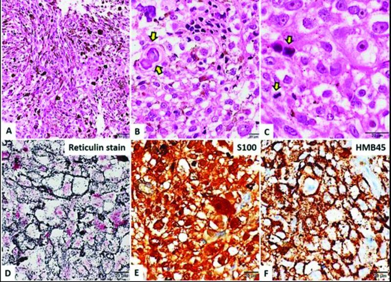
Fig. 2.

( a ) Microphotograph showing a moderately cellular neoplasm composed of spindled to epithelioid cells arranged in interlacing fascicles which are heavily laden with brownish black melanotic pigment. ( b ) Interspersed few psammoma bodies. ( c ) Cells have vesicular nuclei with prominent macronucleoli. Mitotic figures were evident ( arrow ). ( d ) Tumor shows rich pericellular reticulin as highlighted by Gordon and Sweet's method for reticulin fibers. ( e ) Tumor cells were immunoreactive for S100. ( f ) Tumor cells were immunoreactive for HMB45. Magnification as shown in bar.
