Fig. 1.
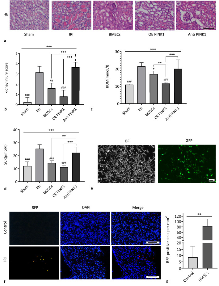
PINK1 enhances BMSC-mediated repair of kidney tissues in IRI-AKI mice. a Representative image of HE staining, × 200, scale: 100 μm. b Pathological score of renal tubular injury (n = 6). c The levels of BUN in blood samples (n = 6). d The levels of SCR in blood samples (n = 6). e BMSCs were successfully transfected with GFP-PINK1, × 100, BAR: 100 μm. f RFP-BMSCs in kidney tissues were observed with a fluorescence microscope. × 200, BAR: 100 μm. g Quantitative analysis of RFP-BMSCs in injured tissues. SEM, ###p < 0.001, ##p < 0.01 and #p < 0.05, compared with the IRI group; ***p < 0.001, **p < 0.01 and *p < 0.05, compared with among the groups

