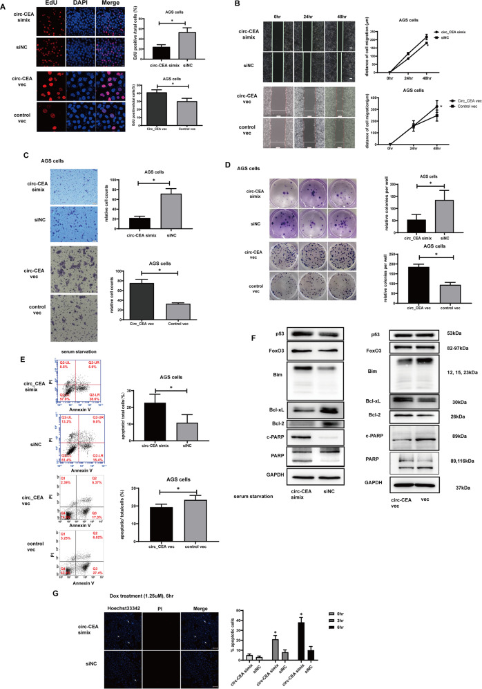
Fig. 2. circ_CEA enhances progression of GC, and suppresses stress-induced apoptosis in GC cells.
A Cellular proliferation was evaluated by EdU assay in AGS transfected with circ_CEA simix or circ_CEA overexpression vector. B Cellular migration was determined via wound-healing assay in AGS cells transfected with circ_CEA simix or circ_CEA overexpression vector. C Cellular migration was further evaluated by transwell migration assay in AGS cells transfected with circ_CEA simix or circ_CEA overexpression vector. D Colony formation assay was performed in AGS cells following the transfection of circ_CEA simix or circ_CEA overexpression vector. E AGS cells was transfected with circ_CEA simix or circ_CEA overexpression vector, followed by serum starvation treatment for 72 hr. The serum starvation-induced apoptosis was evaluated via Annexin V/ PI staining, and apoptotic cells were measured by flow cytometry. F After serum starvation for 72 hr, the expression levels of apoptosis-associated proteins were evaluated by western blotting assay in AGS cells transfected with circ_CEA simix or circ_CEA overexpression vector. G AGS cells were transfected with circ_CEA simix or siNC, followed by Dox treatment (1.25 μM) for the indicated time. The Dox-induced apoptosis was evaluated via hoechst 33342/PI staining. White arrows indicate Hoechst 33342 positive/PI-negative cells (apoptotic cells). *P < 0.05 vs the percentage of apoptotic cells in corresponding siNC-transfected cells. Data are represented as mean± standard deviation (SD).

