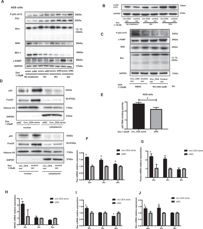
Fig. 4. circ_CEA promotes CDK1-mediated p53 phosphorylation at Ser315 and suppresses p53 activity in GC.
A AGS cells transfected with circ_CEA simix or siNC were treated with Dox (1.25 μM) for the indicated time. The expression levels of apoptosis-associated proteins were evaluated by western blotting assay. B AGS cells transfected with circ_CEA expression vector or control vector were treated with Dox (1.25 μM) for the indicated time, and the level of p53 phosphorylation at ser315 was evaluated by western blotting assay. C AGS cells transfected with circ_CEA expression vector or control vector were treated with Dox (1.25 μM) for 6 hr, in the presence or absence of RO-3306 (2 μM), a selective inhibitor of CDK1. Then the levels of apoptosis-associated proteins were evaluated by western blotting. DMSO was used as a vehicle control for Ro-3306. D AGS cells transfected with circ_CEA simix or siNC were treated with Dox (1.25 μM) for 2 hr (upper). In addition, AGS cells transfected with circ_CEA or control vectors were treated with Dox (1.25 μM) for 2 hr (lower). Then nuclear and cytoplasmic proteins were isolated, respectively. The levels of p53 and FoxO3 in nuclear and cytoplasmic fractions were determined by Western Blotting assay, respectively. Histone H3 and GAPDH were used as positive controls in nuclear and cytoplasmic fractions, respectively. E After Dox treatment (1.25 μM) for 1 hr, p53 DNA binding activity was evaluated in AGS cells transfected with circ_CEA simix or siNC. F–J AGS cells were transfected with circ_CEA simix or siNC, followed by Dox treatment for indicated time. The expression levels of p53 target genes (Fas, Puma, NOXA and Bax) and FoxO3 target gene, Bim were evaluated by qRT-PCR assay. *P < 0.05 vs corresponding siNC-transfected cells. Data are represented as mean± standard deviation (SD).

