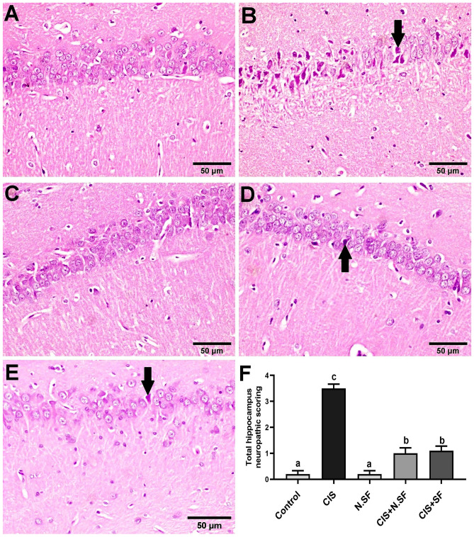
Fig. 11.
Representative photomicrographs of H&E-stained hippocampus of different experimental groups. A Negative control showing the normal histological architecture. B CIS-neurotoxicated brains, showing marked shrunken and pyknosis of pyramidal neurons (arrow). C N.SF-alone treated brains showing no histopathological changes. D and E CIS + N.SF- and CIS + SF-treated brains respectively showing sparse necrosis of pyramidal neurons (arrow) (scale bar: 50 μm). F Total histological scoring of hippocampus damage. Results are presented as mean ± standard error of the mean (SEM). Mean with different letters (a–c) is significant at p ≤ 0.05

