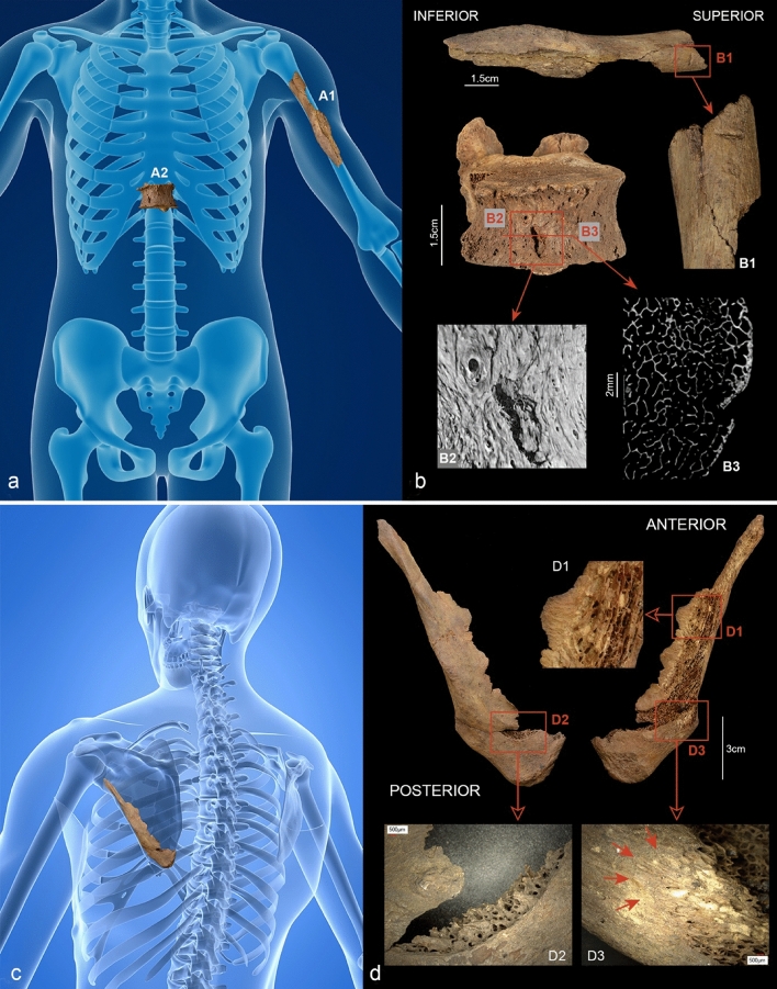
Figure 4.
(a–d) Reconstructed bone injuries. (a and b) The injuries to the left humerus (A1) and the 11th vertebral body (A2). (A1) and (B1) show a kerf in the upper third of the left ventro-lateral humeral diaphysis. The defect, which runs transversely to the longitudinal axis, is 4.9 mm long and shows a V-shaped profile. (A2) and (B2) show the 11th thoracic vertebra which bears a slit-like ventral defect measuring approx. 6 mm in length. The micro-CT images show a terraced depression with small hairline cracks on the left side of the defect (B2); in the transverse section (B3) the lesion in the vertebral body reaches a depth of approx. 3 mm. (c and d) Defect on the left scapula. The bone is dissected by a hard tissue defect approx. 120 mm long with bevelled edges (D1). A crescent-shaped defect is visible above the inferior angle of the scapula (D2). The bone surface appears peeled in some areas (D3) (photos: N. Nicklisch, G. Schulz, (a) Background: A. Ciprian/Shutterstock.com modified by N. Nicklisch, (c) Background: SciePro/Shutterstock.com modified by N. Nicklisch).

