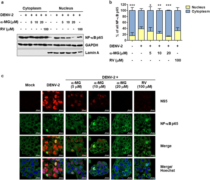
Figure 6.
α-MG inhibits NF-κB nuclear translocation. HepG2 cells were infected with DENV-2 at a MOI of 5 and treated with 5, 10, or 20 μM of α-MG or 100 μM of RV. HepG2 cells that were not infected with DENV-2 were used as mock control. The cells were harvested at 24 h after treatment. Cytoplasmic and nuclear proteins were fractionated. (a) The expression of NF-κB p65 was determined by Western blotting using an anti-NF-κB p65 antibody. NF-κB p65 bands were scanned and quantified using ImageJ software. Lamin A was used as a protein marker of nuclear fraction. (b) The levels of NF-κB p65 were quantified, and the average values from three independent experiments were calculated after being normalized to the level of the GAPDH protein. The intracellular distribution of NF-κB p65 between the nucleus and cytoplasm was calculated from the levels of NF-κB p65 in the nucleus or cytoplasm divided by the total nuclear plus cytoplasmic levels of NF-κB p65. Two-way ANOVA and Bonferroni posttest were used to determine differences of NF-κB p65 levels between nuclear and cytoplasmic fractions and between treatment groups. (c) Immunofluorescence assay demonstrates the expression and intracellular localization of NF-κB p65 (green) and DENV NS5 (red). Hoechst stain was used for nuclear staining (blue). Images are representative of three independent experiments.

