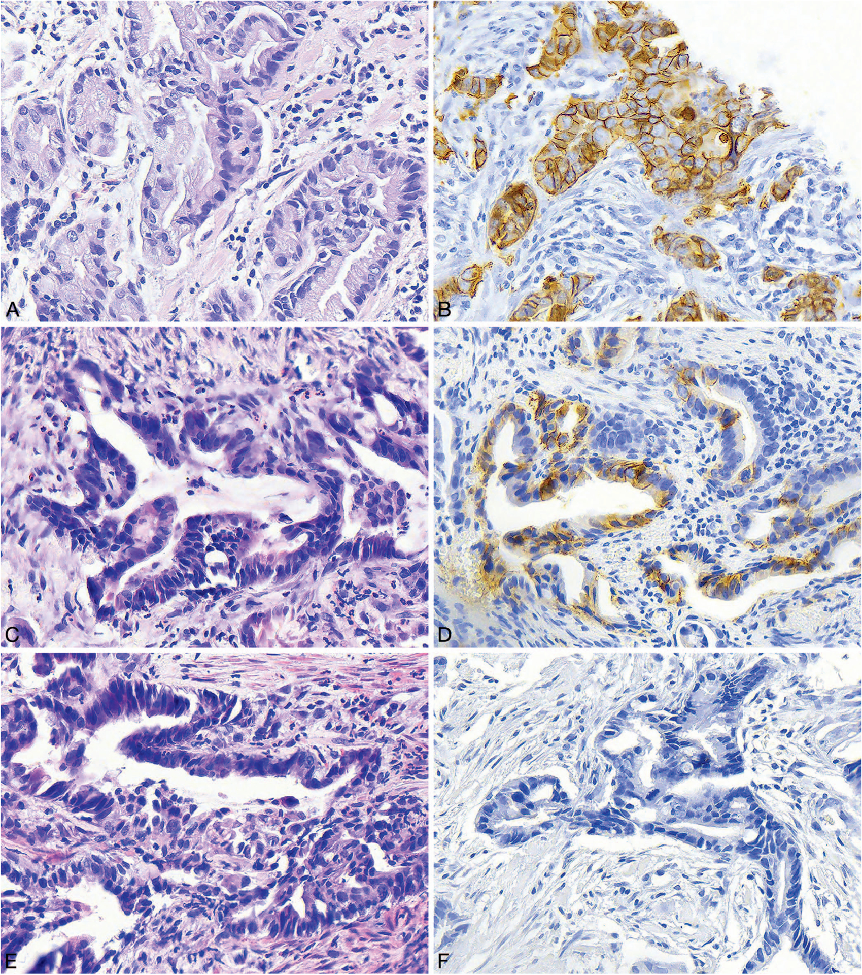
Figure 2.

A and B, Histologic section and human epidermal growth factor receptor 2 (HER2) immunohistochemistry (IHC) from a biopsy specimen showing well-differentiated, intestinal-type gastric adenocarcinoma. The IHC shows malignant glands with strong 3+ staining. C and D, Histologic section and HER2 IHC from a biopsy specimen showing moderately differentiated, intestinal-type gastric adenocarcinoma. The IHC shows malignant glands with equivocal 2+ staining. E and F, Histologic section and HER2 IHC performed on a biopsy specimen showing moderately differentiated, intestinal-type gastric adenocarcinoma. The IHC shows malignant glands with no staining (hematoxylin-eosin, original magnification ×400 [A, C, and E]; HER2 IHC, original magnification ×400 [B, D, and F]).
