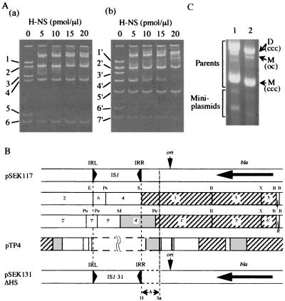
FIG. 3.
(A) Ethidium bromide-stained acrylamide gels (4%) showing the results of the gel retardation assay with H-NS and restriction fragments derived from pSEK117. (a) Restriction fragments digested with BspHI, EcoRI, PstI, SalI, and XmnI and (b) those digested with BspHI, MluI, PvuII, and XmnI. Fragments were incubated with H-NS protein at the indicated concentrations (pmol/μl). Numbers refer to fragments whose positions are shown in panel B. (B) Restriction maps of pSEK117 and pTP4 (a derivative of pUC119 with a cloned fragment [18]). The restriction fragments from pSEK117 are numbered in order of size. Restriction sites: B, BspHI; E, EcoRI; H, HindIII; M, MluI; Ps, PstI; Pu, PvuII; S, SalI; Sa, SapI; X, XmnI. The bla gene is shown with a thick arrow. ori is the replication origin of pUC119. The region in pTP4 which differs from that in pSEK117 is shown by broken lines. An HaeIII restriction map of pTP4 is depicted. The restriction fragments of pSEK117 and pTP4 preferentially bound by H-NS are indicated by hatched or dark boxes. Restriction fragments indicated by the dark boxes required a greater amount of H-NS protein for retardation than those indicated by the hatched boxes. pSEK131ΔHS has a deletion of the HindIII-SapI fragment (dotted lines) which borders the IRR sequence in pSEK117. (C) An ethidium bromide-stained agarose gel (0.7%) showing the production of miniplasmids from pSEK131 (lane 1) and pSEK131ΔHS (lane 2) in YK4122. For further details, see the legend for Fig. 1.

