Figure 3.
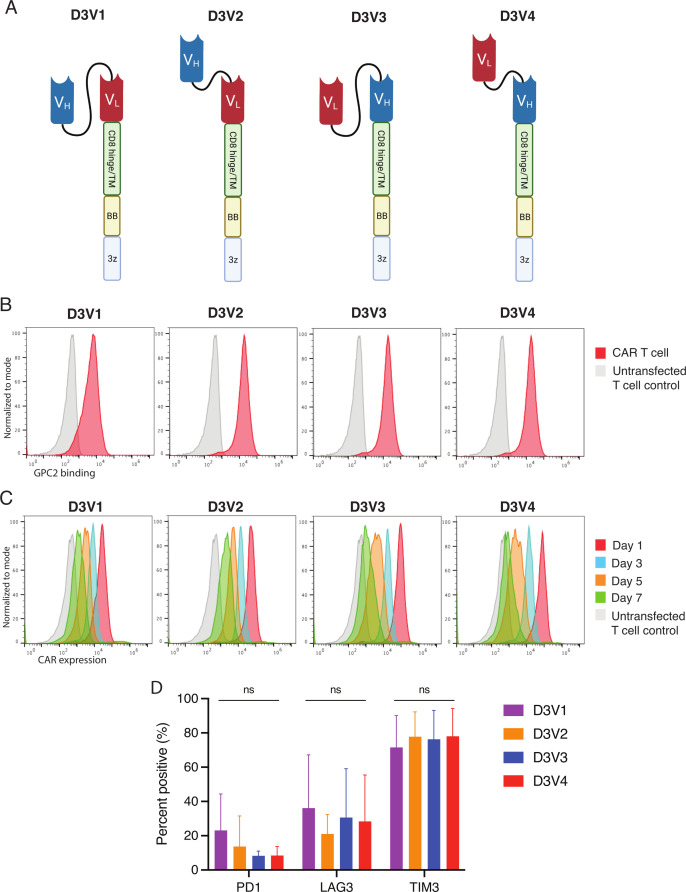
D3-binder based GPC2-directed mRNA CARs specifically bind GPC2 and are transiently expressed on T cells. (A) Schematic of GPC2 CAR constructs. (B) Flow cytometry representative histograms of GPC2-specific binding by GPC2 CAR T cell constructs. T cells were incubated with GPC2 protein conjugated to PE. Red line is CAR T cell, and gray line is untransfected T cell control. MFI is normalized to mode. (C) Flow cytometry representative histograms of GPC2 CAR persistence on T cells. T cells were incubated with protein L to identify CAR expression. Red line represents day 1 post-transfection, blue line represents day 3, orange line day 5, and green line day 7. Gray line is untransfected T cell control. Mean fluorescence intensity (MFI) is normalized to mode. (D) Cell surface expression of negative checkpoint regulators PD1, Lag3, and Tim3 quantified with flow cytometry at day 4 post-transfection. Data are represented as mean±SD of five independent experiments. Graphics in figure part A created with BioRender.com. 3z, CD3ζ co-stimulatory domain; BB, 41-BB costimulatory domain; CARs, chimeric antigen receptors; GPC2, glypican 2; ns, not significant; PE, phycoerythrin; TM, transmembrane domain; VH, variable heavy chain; VL, variable light chain.

