
Fig. 4.
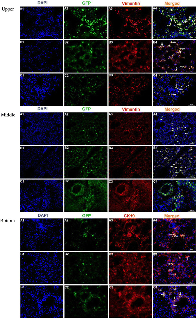
Upper: GFP labeled rBM-MSCs in rat lung tissue after transplantation. rBM-MSCs were labeled with GFP and injected intratracheally into rats. Immunofluorescence of paraffin lung sections examined by flouresans microscopy revealing colocalization of BM-MSCs (green) and the MSCs marker vimentin (red) together with nuclear staining (4′,6-diamidino-2-phenylindole [DAPI], blue). GFP+ MSCs (green) were located in perivascular, peribronchial and interalveolar area (indicated by arrows) (A4–C4). Those cells were also positive for MSC marker, vimentin (red) (Scale Bars: 100 μm). Middle: Localization of GFP + rBM-MSCs in lung tissue samples of different rats. GFP+ cells (white arrows; green fluorescence) could be observed in the interalveolar septae (A2, B2, C2, A4, B4, C4). The labeled cells (white arrows) could also be located in both peribronchial and perivascular areas of the lung. Some of these cells (red arrow) were observed positive for vimentin (red fluorescence) (C4) (Scale bars: 100 μm). Bottom: Immunocytochemical expression patterns for GFP (green) and CK19 (red) were colocalized in the cytoplasm of epithelial cells (visualized as orange/yellow fluorescence in merge panel). These GFP+ cells had not only acquired proper epithelial morphology, but it also stained positive for specific antigens such as CK19. In addition, specific GFP+ cells were observed within bronchiolar epithelial cells (Scale bars: 100 μm)