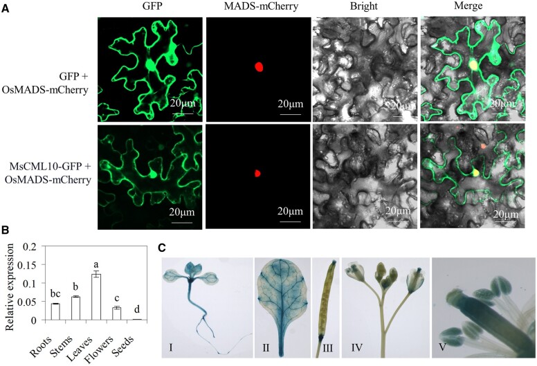
Figure 1.
Subcellular localization of MsCML10 and the expression pattern. The vectors MsCML10::GFP or GFP in combination with mCherry-tagged MADS were co-transformed into N. benthamiana leaves for analysis of subcellular localization (A). Spatial expression of MsCML10 in alfalfa was analyzed using RT–qPCR (B). Seedlings (I), leaflet (II), silique (III), flower (IV), and stamen (V) in PMsCML10::GUS transgenic Arabidopsis was used for GUS staining (C). Means of three replicates and standard errors are presented; the same letter above the column indicates no significant difference at P < 0.05 using Duncan’s test.

