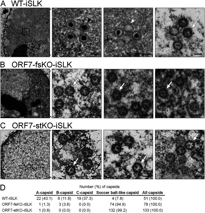
FIG 1.
TEM images showing capsids produced from lytic-induced WT-KSHV- and ORF7-KO-KSHV-harboring iSLK cells. iSLK cells harboring WT or ORF7-KO KSHV were cultured for 72 h in medium containing Dox and NaB to induce the lytic phase. Capsid formation in nuclear (A) WT-iSLK, (B) frameshift-induced ORF7 KO (ORF7-fsKO-iSLK), and (C) stop codon-induced ORF7 KO (ORF7-stKO-iSLK) cells as observed by TEM. The black and white arrowheads indicate C-capsids and A-capsids, respectively, and the white arrows indicate soccer ball-like capsids. The scale bars represent a length of 100 nm. (D) Quantification of the number of each type of capsid observed by TEM.

