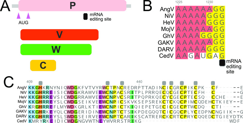
FIG 3.
Organization of the P gene of AngV. (A) Alternative transcriptional start sites (pink triangle) generate the P and C protein. Pseudotemplated addition of one or two guanine nucleotides at the putative mRNA editing site generates a V and W protein, respectively. (B) Sequence alignment of the putative mRNA editing site across members of the Henipavirus genus (cRNA depicted). (C) Amino acid alignment of the unique C-terminal region of the V protein following the addition of one guanine nucleotide to the putative mRNA editing site. Gray boxes denote conserved cysteine and histidine residues suggested to directly coordinate bound zinc ions (32). Individual nucleotides or amino acids are color coordinated if at least 75% conserved at the alignment position. Nucleotide or amino acid position numbers displayed represent the position within the AngV gene or protein. Virus name (abbreviation), followed by GenBank accession number: Angavokely virus (AngV) ON613535; Nipah virus (NiV) AF212302; Hendra virus (HeV) AF017149; Mojiang virus (MojV) KF278639; Ghanaian bat Henipavirus (GhV) HQ660129; Daeryong virus (DARV) MZ574409; Gamak virus (GAKV) MZ574407; Cedar virus (CedV) JQ001776. CedV is shown here only for comparison, as the CedV P protein is not believed to undergo RNA editing or to generate a functional V protein (8, 32).

