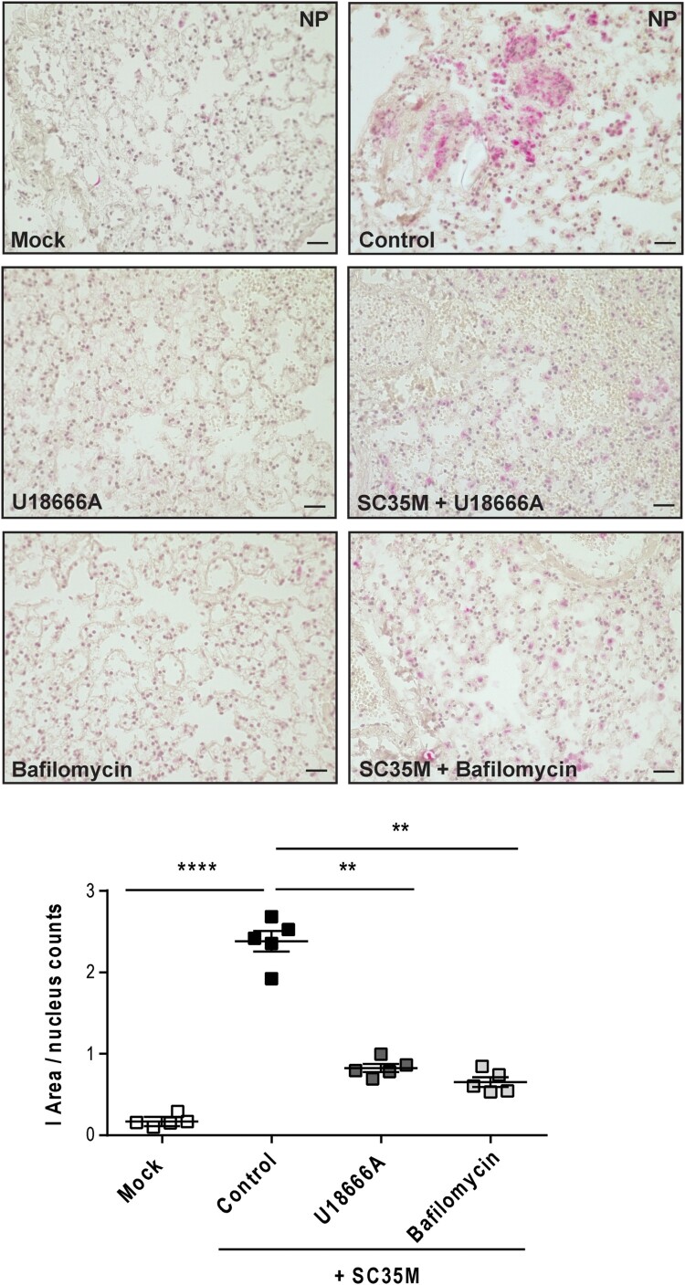
Figure 4.
Histological evaluation of the IAV infection rate. Murine lungs were either pre-treated with the solvent DMSO (control), 250 nM bafilomycin A (1 h) or with 10 µg/mL U18666A (16 h) prior to infection, followed by an infection with 105 PFU/ mL of IAV strain SC35M for 24 h. Representative images of NP immunostaining detected in the lung sections. Quantitative analysis of NP staining. Scatter plot representation of individual lungs, with means ± SEM superimposed. Data were analyzed by Kruskal-Wallis test followed by Dunn's multiple comparisons test, * p < 0.1, ** p < 0.01, **** p < 0.0001, n = 5 murine lungs/group, scale bar 50 µm.

