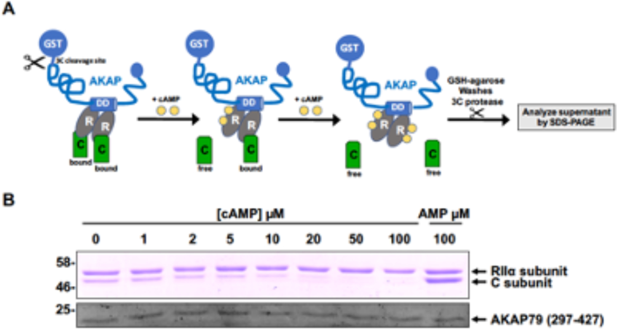
Fig. 3.

Biochemical analysis of the AKAP:RII:C interaction in the presence and absence of exogenous cAMP titrated over a range of concentrations. (A) Cartoon of the assay procedure, which detects free C-subunits released into solution from previously assembled AKAP:RII complexes (B) SDS-PAGE showing the electrophoretic mobility and staining intensity of 3C-generated cleaved AKAP and the relative amounts of associated RII and C subunits. The amount of 3C-cleaved AKAP and RII remain constant in the assay. A high concentration of 5’AMP serves as an internal control, since it does not lead to C-subunit release.
