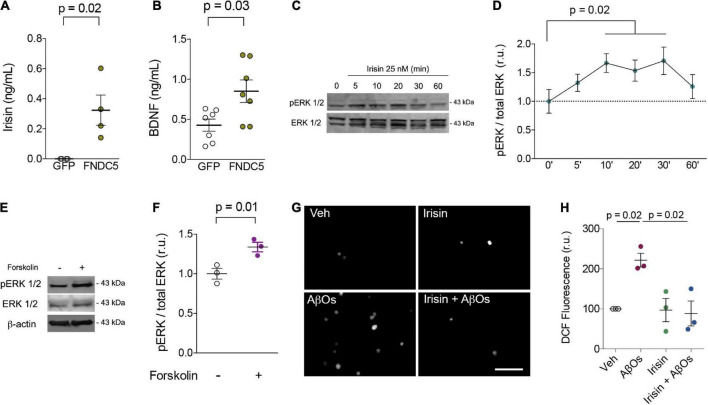
FIGURE 1.
Irisin increases extracellular BDNF levels, stimulates transient ERK activation, prevents AβO-induced oxidative stress in primary neurons. (A,B) Primary hippocampal cultures were transduced with AdFNDC5 or AdGFP (control) adenoviral particles for 48 h. Conditioned media were collected and levels of irisin (A) or BDNF (B) were assessed by ELISA. N = 4 experiments using independent hippocampal cultures for irisin and 7 for BDNF measurements. Wilcoxon matched-pairs rank test. Primary hippocampal cultures were treated with recombinant irisin (25 nM) for the indicated timespoints (C,D) or with forskolin (10 μM; 20 min) (E,F), and ERK 1/2 phosphorylation at Thr202/Tyr204 (pERK 1/2) was measured by immunoblotting. N = 3 experiments using independent hippocampal cultures. Repeated measures one-way ANOVA with Holm-Sidak correction. (G,H) Hippocampal neurons were exposed to 0.5 μM AβOs for 3 h in the presence or absence of recombinant irisin (25 nM). When present, irisin was added 15 min before AβOs. ROS was measured by DCF fluorescence. N = 3 experiments with independent cultures and AβO preparations. Two-tailed two-way ANOVA with Holm-Sidak correction. Each dot represents an independent hippocampal culture; data are shown as means ± S.E.M. p-values are indicated in the figure. Scale bar = 100 μm.

