Figure 2.
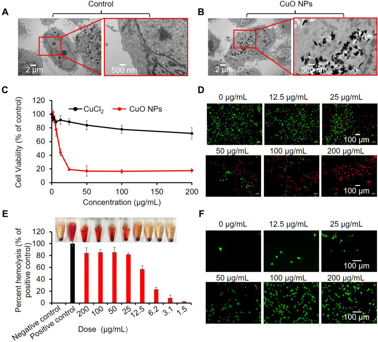
Cytotoxic properties of CuO NPs. (A) TEM images of BEAS-2B cells before exposure and (B) after exposure to 25 μg/mL CuO NPs for 6 h. (C) Cell viability assessment of Cu ions (CuCl2) and CuO NPs in BEAS-2B cells. Cells were exposed to serial concentrations (0, 0.4, 0.8, 1.5, 3.1, 6.25, 12.5, 25, 50, 100, and 200 μg/mL) of Cu ions or CuO NPs, and cell viability was assessed by the CCK-8 assay. (D) Live/dead staining of BEAS-2B cells treated with CuO NPs at 200, 100, 50, 25, 12.5 and 0 μg/mL for 24 h. The live and dead cells were stained with calcein AM (green) and PI (red), respectively. (E) Hemolytic effect of CuO NPs. Mouse RBCs were exposed to CuO NPs for 3 h. Inset picture showing hemoglobin appearance (red) in the RBC solution. (F) CLSM images of BEAS-2B cells reflecting intracellular ROS (DCF, green).

