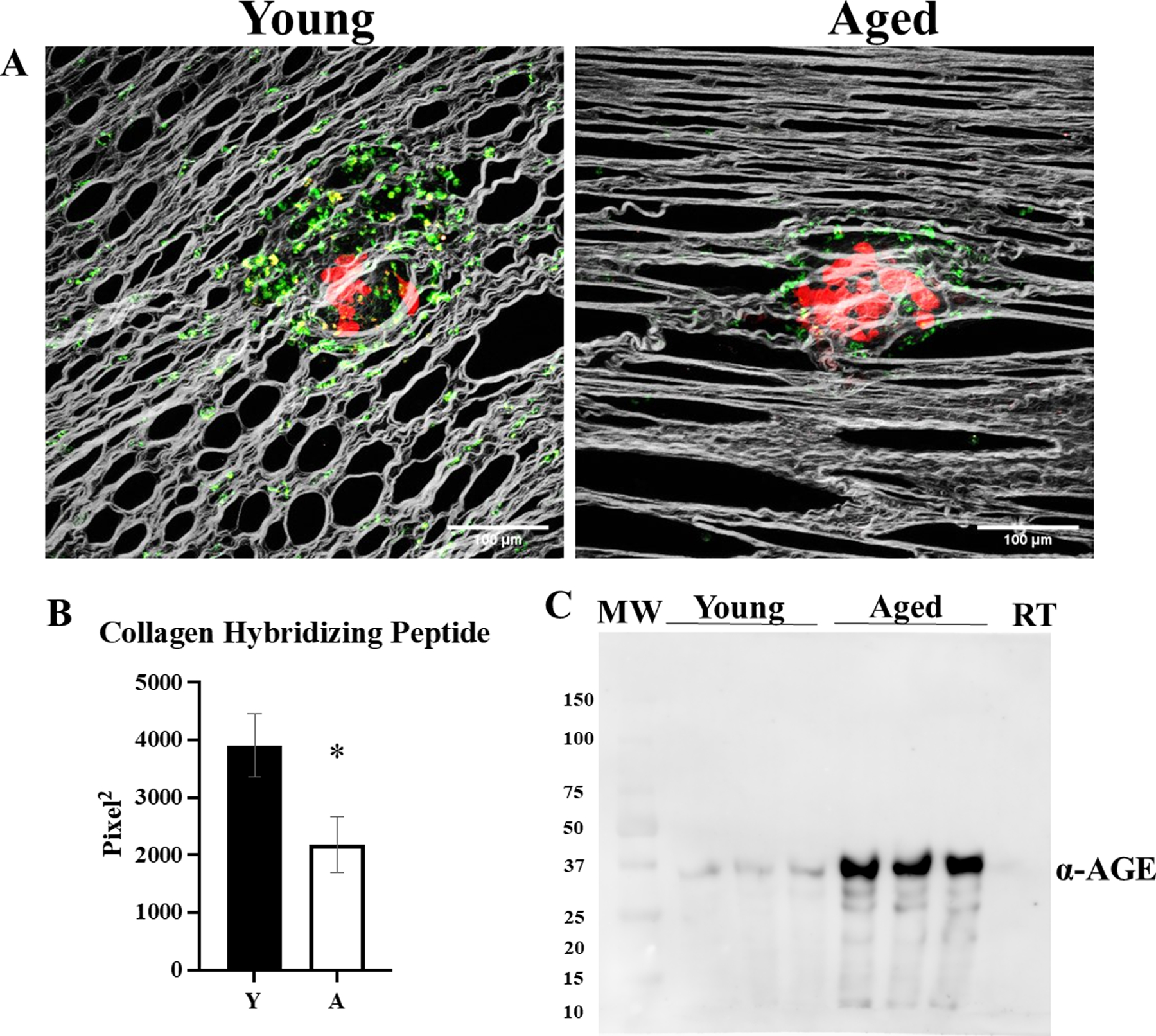
Figure 5. Visualization of peri-tumoral collagen remodeling in young and aged mice.

C57Bl/6 mice (n=5 per cohort) at 3–6 months of age (Young; Y) or 20–23 months of age (Aged; A) were injected IP with RFP-tagged syngeneic ID8trp53−/− cells (107). Tumors were allowed to grow for 3 weeks, then mice were injected with CHP-B:Streptavidin-Alexa Fluor 647 and sacrificed 3 hr post injection and omenta were imaged with SHG microscopy in conjunction with 2-photon excitation fluorescence microscopy to (A) visualize collagen (grey), tumor cells (red), and biotin conjugate of the collagen hybridizing peptide bound to Alexa Fluor 647 conjugated to streptavidin (pseudocolored green). (B) The images were analyzed for collagen hybridizing peptide signal (p=0.04). (C) Collagen isolated from young and aged mice, as well as commercially available rat tail (RT) collagen, was electrophoresed on a 9% SDS-PAGE gel, transferred to a PVDF membrane, probed with anti-AGE (1:500) and developed with a peroxidase-conjugated secondary antibody (anti-rabbit, 1:4000) and ECL detection. Error bars represent standard error of mean and p-values were determined by Student’s t-test.
