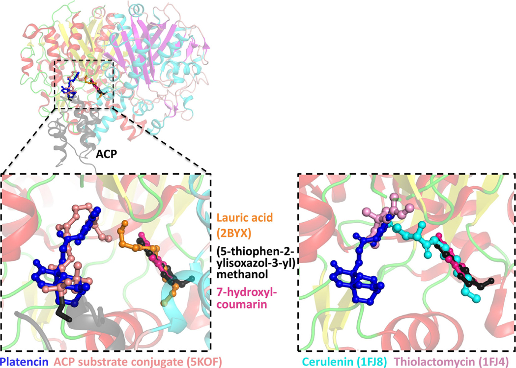
FIGURE 7.
Left panel: the structure of the ACP substrate conjugate (5KOF) (colored light pink) and lauric acid (2BYX) (colored orange) bound to β-ketoacyl-ACP synthase I were superimposed onto the structures determined in this study. Platencin (dark blue) bound in the same site as the ACP substrate conjugate, while 7-hydroxyl-coumarin (dark pink) and (5-thiophen-2-ylisoxazol-3-yl)methanol (black) superimposed with lauric acid. Right panel: Cerulenin (cyan) (1FJ8) and thiolactomycin (pink) (1FJ4) also superimposed with the ligands determined in this study

