
Figure 3.
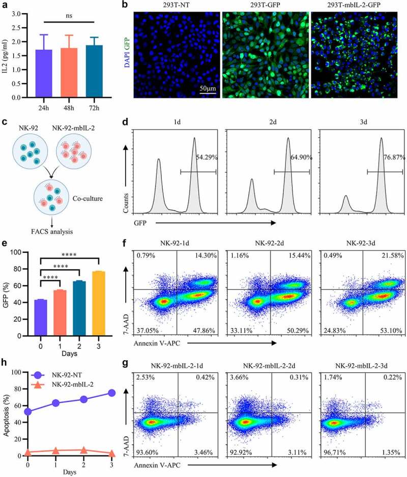
MbIL-2 does not support bystander cell survival and proliferation.
a: ELISA detects IL-2 in NK-92-mbIL-2 cell culture supernatants at different times (n = 3). NK-92-mbIL-2 maintained without IL-2 and collected culture supernatants for assays. b: Representative confocal laser images of 293 T-NT, 293 T-GFP, and 293 T-mbIL-2-GFP. 293 T-NT represent 293 T cells not-transduced lentiviral. 293 T-GFP represent 293 T cells transduced empty lentiviral carrying GFP reporter gene. 293 T-mbIL-2-GFP represent 293 T cells transduced lentiviral carrying mbIL-2-GFP fusion protein. c: Schematic diagram of NK-92-mbIL-2 cell co-culture with NK-92 cells. At the co-culture time, IL-2 was not added. d: Representative flow cytometry analysis showing the proportion of NK-92-mbIL-2 in co-culture assays shown by C. GFP positive cells represent NK-92-mbIL-2 cells which carrying GFP reporter gene. GFP negative cells represent non-transduced NK-92 cells. e: Quantification and statistical analysis of the data in D (n = 3). f: Representative flow cytometry analysis showing the proportion of apoptotic non-transduced NK-92 cells in co-culture assays shown in C. g: Representative flow cytometry analysis showing the proportion of apoptotic NK-92-mbIL-2 cells in co-culture assays shown in C. h: Quantification and statistical analysis of the data in F and G (n = 3).