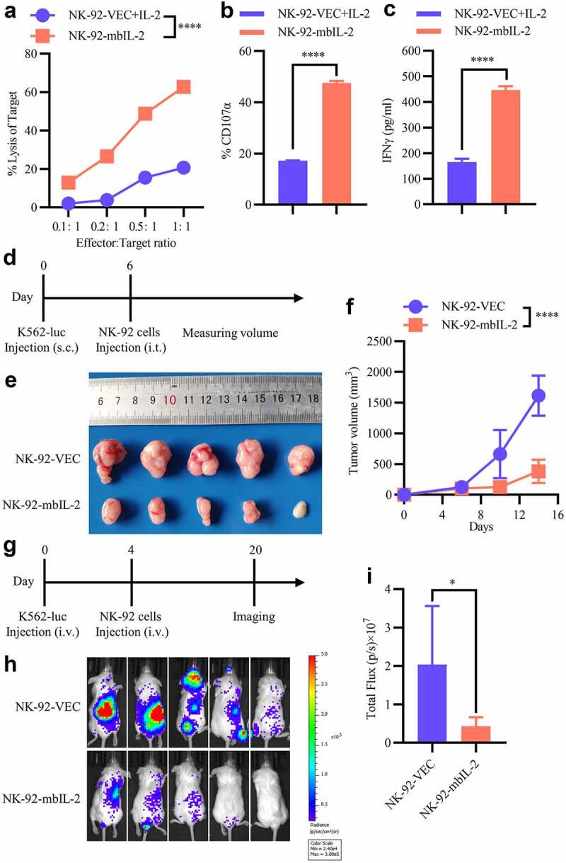
Figure 4.

MbIL-2 enhances the antitumor activity of NK-92 cells.
a: Direct lysis of NK cells against target cells K562. Effector and target cells were co-incubated for 4 h at the indicated effector: target ratio. Flow cytometric analysis of the proportion of GFP−7-AAD+ cells (n = 3). b: Flow cytometry analysis of the proportion of GFP+-CD107α+ cells after NK cells were co-incubated with target cells at 1:1 for 5 h (n = 3). c: ELISA data showing the release of IFN-γ by NK cells after co-incubation with target cells at 1:1 for 12 h (n = 3). d–f: In vivo studies using K562-luc cells in a mouse subcutaneous xenograft model treated with NK-92-VEC and NK-92-mbIL-2 cells. d: Schematic diagram of the study. 1 × 10⁶ K562-luc cells with Matrigel Matrix were subcutaneously injected to establish a xenograft model. After the volume of the tumors reached 100–200 mm3, 5 × 10⁶ NK-92-VEC or NK-92-mbIL-2 cells were injected intratumorally (n = 5). Subsequently, the volume of the tumors was measured periodically. e: Representative image of tumor burden after the mice were sacrificed (n = 5). f: Tumor burden was periodically determined (n = 5). g–i: In vivo study using K562-luc cells in a mouse orthotopic xenograft model treated with NK-92-VEC and NK-92-mbIL-2 cells. g: Schematic diagram of the study. 1 × 10⁶ K562-luc cells were injected intravenously to establish an in-situ model of leukemia. After four days, 5 × 10⁶ NK-92-VEC or NK-92-mbIL-2 cells were injected intravenously (n = 5). Tumor burden was measured by in vivo bioluminescence using the Xenogen-IVIS Imaging System. h: Representative bioluminescent image of the tumor burden after mice were treated (n = 5). i: Quantification and statistical analysis of the data in H (n = 5).
