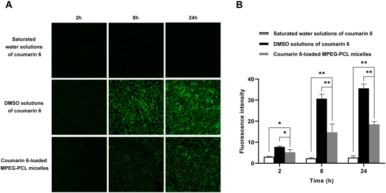
Figure 5.
In vitro cellular uptake of MPEG-PCL micelles. (A) Fluorescence images of HUVEC uptake of coumarin 6-loaded MPEG-PCL micelles, saturated water solutions and DMSO solutions of coumarin 6. (B) Quantification of the fluorescence intensity of coumarin 6 in HUVECs. Data represent the mean±SD, n=3. *P < 0.05, **P < 0.01.

