
Figure 4.
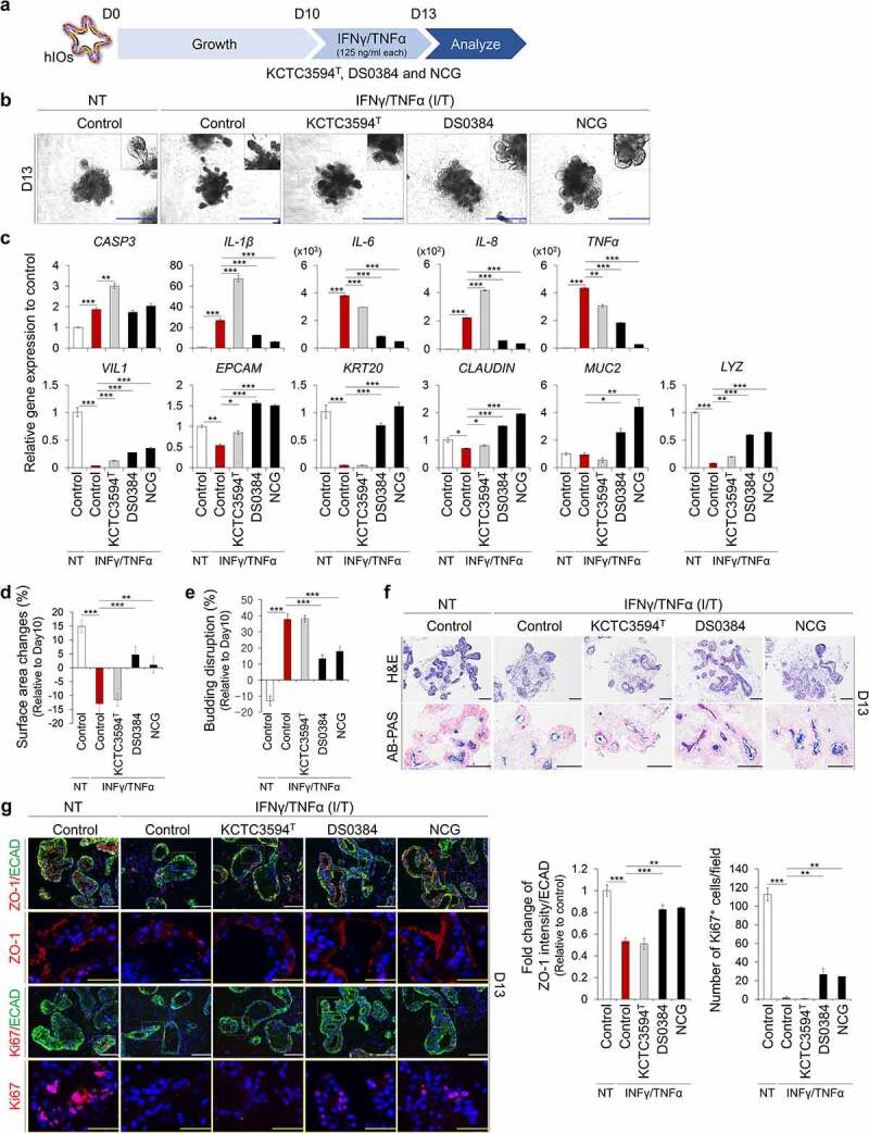
Protective effects of postbiotic metabolites produced by L. reuteri DS0384 on IFNγ/TNFα-induced inflamed hIO. (a) Schematic diagram of experimental design. (b) Morphology of control and IFNγ/TNFα-induced inflamed hIOs following treatment with cell-free supernatants (CFS) from L. reuteri KCTC3594T and DS0384, and metabolite NCG. Scale bar, 1 mm. (c) qPCR analysis of apoptosis, inflammatory, intestine-specific, and intestinal barrier markers in inflamed hIOs and control hIOs (n > 10) treated with CFS from L.reuteri KCTC3594T and DS0384, and metabolite NCG. (d) Percentages of surface area changes. Data represent the means ± SEM (n > 10). (e) Percentages of budding disruption. Data represent the means ± SEM (n > 10). (f) Histological analysis of control and inflamed hIOs following treatment with CFS from L. reuteri DS0384. Scale bar, 200 μm. (g) Immunofluorescence analysis of intestinal barrier (zonula occludens-1, ZO-1) and proliferating marker (Ki67). White scale bar, 125 μm, yellow scale bar, 50 μm. Fold-change in intestinal barrier (ZO-1) intensity to intestinal epithelial marker (ECAD) (middle, n > 5). Number of Ki67+ cells per field (right), (n = 5–10 10x fields from at least 10 organoids). Data are the mean ± SEM. *p < .05, **p < .01, ***p < .001 by two-tailed t-test.