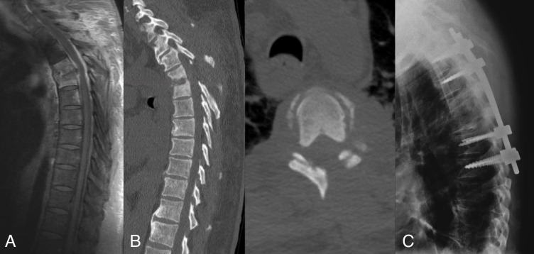
Figure 2.
(A) Sagittal magnetic resonance imaging with contrast of the thoracic spine in a patient who presented with upper thoracic back pain showing discitis-osteomyelitis at T4-5. (B) Sagittal and axial postoperative computed tomography images showing a left-sided T4-5 hemilaminectomy, which was the initial surgical treatment for this patient. (C) Sagittal x-ray images showing the patient’s reoperation, having underwent a T4-5 redo laminectomy and a T2-7 posterior fusion after the patient presented with persistent mechanical back pain, especially with axial loading.

