
Figure 4.
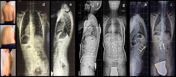
Clinical presentation (a–c) and preoperative x-ray images (d, e); postoperative x-ray images with cast (f, g); and 6 months follow-up x-ray images showing proximal junctional kyphosis (h, i).
Official websites use .gov
A
.gov website belongs to an official
government organization in the United States.
Secure .gov websites use HTTPS
A lock (
) or https:// means you've safely
connected to the .gov website. Share sensitive
information only on official, secure websites.
Clinical presentation (a–c) and preoperative x-ray images (d, e); postoperative x-ray images with cast (f, g); and 6 months follow-up x-ray images showing proximal junctional kyphosis (h, i).