
Figure 5.
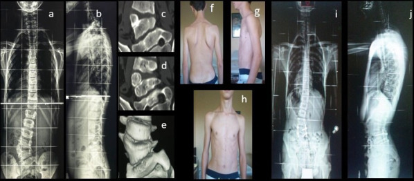
Preoperative x-ray images (a, b), computed tomographic (CT) images (c, d), and three-dimensional CT image (e); and clinical (f–h) and radiologic (i, j) findings at 8 months follow-up, showing small upper scoliotic curve development and slight cosmetic impairment.