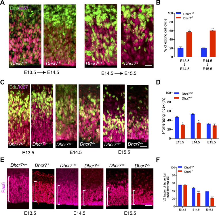
Figure 6. Dhcr7-/- mice show accelerated cell cycle exiting and depletion of cortical precursor cells in vivo.
(A) Cortical sections from Dhcr7-/- and Dhcr7+/+ embryos labeled by Edu at different developmental stages were immunostained 18 hr later for Edu (green) and Ki67 (Magenta). (B) Quantification of cell-cycle exit index of Dhcr7-/- cortices compared with Dhcr7+/+ cortices. (C) Cortical sections from Dhcr7-/- and Dhcr7+/+ embryos labeled by Edu injection at different developmental stages were immunostained 2 hr later for Edu (green) and Ki67 (Magenta). (D) Quantification of proliferation index of Dhcr7-/- cortices compared with Dhcr7+/+ cortices. (E) Coronal cortical sections immunostained for Pax6 cortical precursor marker at different developmental stages. (F) Quantification of the relative size of the Pax6 +region shown as fractions of the whole cortical radial thickness. Error bars indicate SEM. *, p<0.05; **, p<0.005; ***, p<0.001. n=3 per experiment. Scale Bar = 50 μm.

