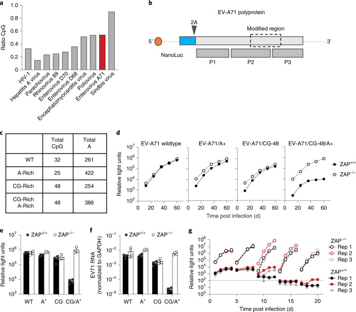
Fig. 4. Genomic recoding sensitizes EV-A71 to ZAP.
a, CpG content of example virus genomes; observed/expected ratios (based on mononucleotide composition) for HIV-1, Sindbis virus and several picornaviruses. b, Schematic diagram of reporter EV-A71 containing the NanoLuc luciferase gene (blue) and a 2A cleavage site. P1, P2 and P3 indicate the primary processed proteins derived from the EV-A71 polyprotein. The recoded region (dashed line box) is approximately 1 kb and encodes a portion of the polyprotein located in P2 and P3. c, Summary of the recoding modifications introduced in the EV-A71 genome. d–f, Replication of reporter EV-A71 mutants in ZAP+/+ or ZAP−/− HeLa cells. NanoLuc luciferase activity was measured every 12 h (d). Summary of NanoLuc luciferase levels at 60 h post infection, mean ± s.d. of 3 independent experiments (e). Viral RNA levels measured at 60 h post infection, mean ± s.d. of 3 independent experiments (f). g, Replication of EV-A71(CG-48)/A+ in ZAP+/+ or ZAP−/− HeLa cells over 4 passages. At 4 d post initial infection, supernatants were collected, filtered through a 0.22 µm filter and used to infect a fresh culture of cells.

