Fig. 1.
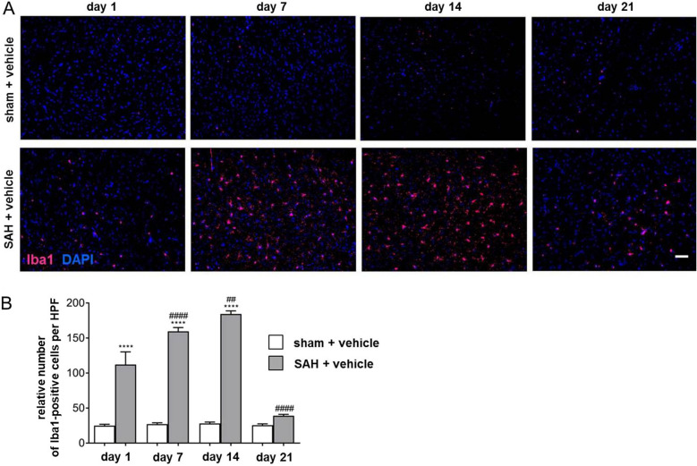
Time-dependent increase of microglia/macrophage accumulation until day 14 after SAH. a Representative images of coronal brain sections of sham + vehicle (upper panel) and SAH-operated mice (lower panel) were chosen to demonstrate the time-dependent accumulation of microglia/macrophages in response to the bleeding. Microglia/macrophages were stained using Iba1 (magenta) and counterstained with DAPI (blue) to visualize cell nuclei. The number of Iba1-positive cells in SAH and sham + vehicle (not provided) was counted using ImageJ software. Bar indicates 60 µm. Microglia/macrophage accumulation was significantly increased in SAH + vehicle-treated mice at all experimental time points compared with the respective sham. b Microglia/macrophage accumulation culminated 14 days after SAH and was reduced at day 21 after the bleeding when compared with the amount counted at day 14, as depicted. Values from the graph are means ± SEM (n = 6 animals per group). ****/####P < 0.0001, ***/###P < 0.001, **/##P < 0.01, and */#P < 0.05 versus sham + vehicle and SAH + vehicle, respectively. Statistical significance was determined by one-way ANOVA Bonferroni corrected. (*) describes significance with respect to sham + vehicle; (#) describes significance with respect to the earlier experimental time point. ANOVA analysis of variance, DAPI 4',6-diamidino-2-phenylindol, Iba1 ionized calcium-binding adapter molecule 1, SAH subarachnoid hemorrhage, SEM standard error of mean (Color figure online)

