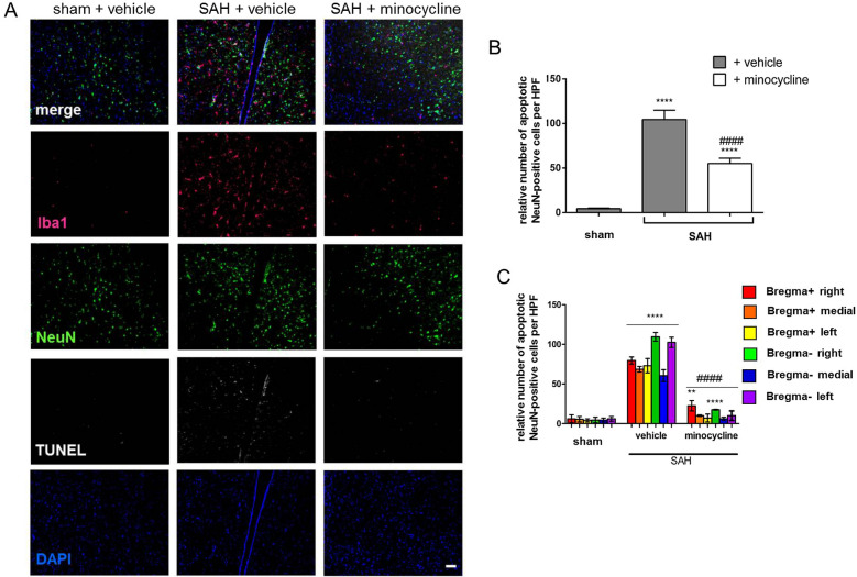
Fig. 5.
SAH-induced neuronal cell death was significantly lowered on minocycline administration. a Mice were operated and treated as indicated. b TUNEL-positive (white) NeuN-labeled and DAPI-labeled (blue) neuronal cell bodies (green) were counted, and the results were summarized. Microglia/macrophages were stained by Iba1. c Regional distribution of TUNEL-NeuN-DAPI double-triple-positive neurons is depicted. Coronal brain sections were analyzed in the area of corpus callosum (bregma +) and hippocampus (bregma −), as indicated by colored squares in Fig. 2c. Bar indicates 20 µm. Values from all graphs are means ± SEM (n = 6 animals per group). ****/####P < 0.0001 versus sham + vehicle and SAH + vehicle, respectively. Statistical significance was determined by one-way ANOVA Bonferroni corrected. (*) describes significance with respect to sham; (#) describes significance with respect to the SAH + vehicle. ANOVA analysis of variance, DAPI 4',6-diamidino-2-phenylindol, Iba1 ionized calcium-binding adapter molecule 1, SAH subarachnoid hemorrhage, SEM standard error of mean, TUNEL terminal deoxynucleotidyl transferase deoxyuridine triphosphate nick end labeling (Color figure online)

