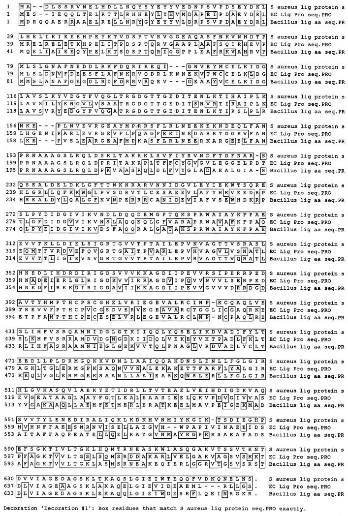
FIG. 1.
Clustal amino acid sequence alignment of DNA ligases from S. aureus, E. coli, and B. stearothermophilus. Regions enclosed in boxes indicate identical amino acid residues. Amino acid positions of conserved motifs (designations from references 4, 8, 9, and 26) for the S. aureus DNA ligase are as follows: motif I, 112 to 117; motif II, 278 to 283; motif III, 190 to 214; and motif IV, 591 to 667.

