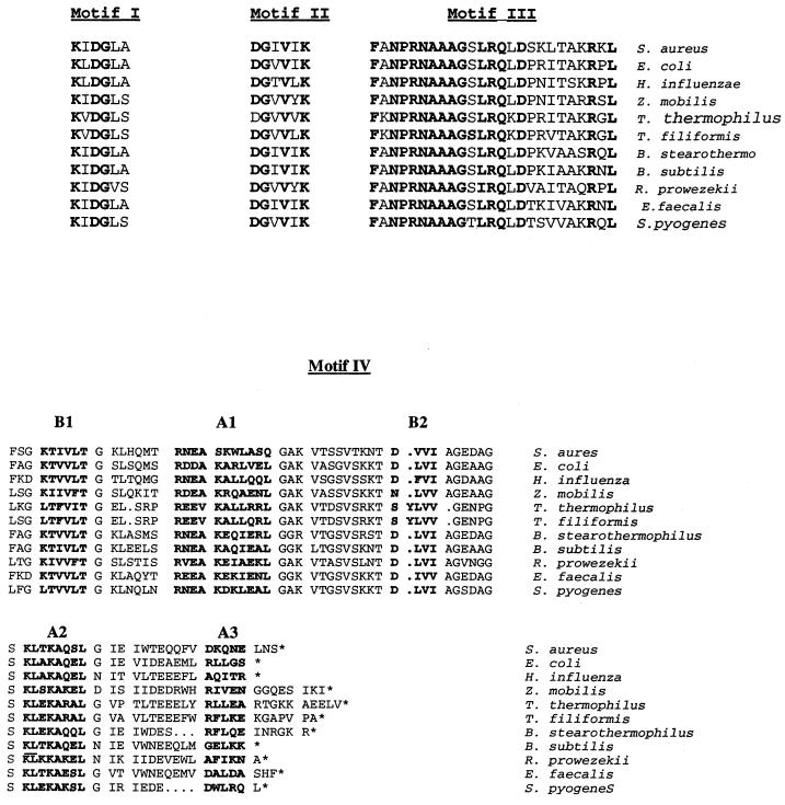
FIG. 2.
Comparison of the conserved amino acid motifs I through IV from known and uncharacterized bacterial ligA gene sequences. Amino acids identical in all species are shown in boldface type. Motif I is the conserved region at the active site. Motifs II and III are of unknown function. Motif IV constitutes the DNA binding domain. Designations in motif IV (B1, A1, etc) refer to areas defined as beta-sheet or alpha-helix structures (4). Alignment of amino acid sequences within the C-terminal DNA binding motif IV from various DNA ligases were as follows: E. coli, aa 598 to 671; Haemophilus influenzae, aa 605 to 679; Zymomonas mobilis, aa 649 to 731; T. thermophilus, aa 594 to 676; T. filiformis, aa 594 to 667; B. stearothermophilus, aa 594 to 670; Bacillus subtilis, aa 595 to 668; Rickettsia prowazekii, aa 619 to 689; S. aureus, aa 591 to 667; Enterococcus faecalis predicted DNA ligase, aa 586 to 664; and Streptococcus pyogenes predicted DNA ligase, aa 582 to 652. The unpublished genomic sequence data used to identify the LigA orthologues were obtained from the PEDANT database (http://pedant.mips.biochem.mpg.de/).

