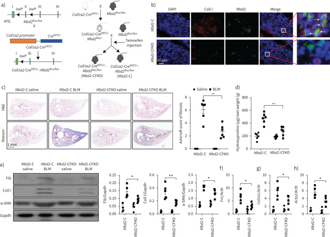
FIGURE 4.
Comparison of the severity of lung fibrosis between methyl-CpG-binding domain 2 (Mbd2)-CFKO and Mbd2-C mice after bleomycin (BLM) induction. a) Mbd2flox/flox mice were generated by inserting two loxP sequences in the same direction into the introns flanked with the exon 2 of MBD2 based on the clustered regularly interspaced short palindromic repeats (CRISPR)–Cas9 system, which could produce a nonfunctional Mbd2 protein by generating a stop codon in exon 3 after Cre-mediated gene deletion. Mbd2flox/flox then crossed with the Coll1α2-CreERT2+ transgenic mice to get the fibroblast-specific Mbd2-knockout mice following intraperitoneal injection of tamoxifen for five consecutive days. b) Representative results for co-immunostaining of Coll I and Mbd2 in lung sections from Mbd2-C and Mbd2-CFKO mice. Nuclei were stained blue using 4′,6-diamidino-2-phenylindole (DAPI), and the images were taken at an original magnification of ×400. c) Histological analysis of the severity of lung fibrosis in mice after BLM induction. Left: representative images for haematoxylin and eosin (H&E) and Masson staining. Right: quantitative mean score of the severity of fibrosis. d) Quantification of hydroxyproline contents in Mbd2-CFKO and Mbd2-C mice after BLM challenge. e) Western blot analysis of levels of fibronectin (Fib), collagen (Coll) I and α-smooth muscle actin (SMA). f–h) Results for reverse transcriptase (RT)-PCR analysis of f) Fn1, g) Coll1a1 and h) Acta2. Five to six mice were included in each study group. Gapdh: glyceraldehyde 3-phosphate dehydrogenase. Data are presented as mean±sd. *: p<0.05, **: p<0.01.

