. 2022 Sep 6;63(10):966–970. doi: 10.3349/ymj.2022.0038

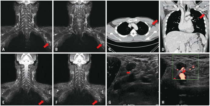
Fig. 2. Imaging findings of the patient. (A and B) Brachial plexus MRI on the fifth day after the onset of symptoms shows multiple enlarged LNs on the left axilla (arrow). There was no evidence of direct invasion to the brachial plexus. The largest LN was approximately 2.5–3.0 cm in length. (C and D) Chest CT performed on the seventh day after the onset of symptoms also reveals multiple enlarged LNs, suggesting the possibility of reactive lymphadenopathy. The size of the largest LN was reduced to 6–7 mm after intravenous steroid loading treatment (arrow). (E and F) MRI for brachial plexus on the 28th day after the onset of symptoms. Compared to a previous study, the size and number of enlarged benign LNs along the carotid and axillary vessels (left>right) have almost normalized (arrow). (G and H) Ultrasonography (USG) on the left axilla level I. USG on the ninth day after the onset of symptoms shows that the biggest LN measures approximately 10.7×7.5 mm in size. Hypoechoic radial nerve is seen compressed by enlarged LN within mixed echogenic soft tissue in the longitudinal plane (arrow). The results of fine needle aspiration suggested benign reactive hyperplasia of the LN. LN, lymph node.

Fig. 2