Figure 1.
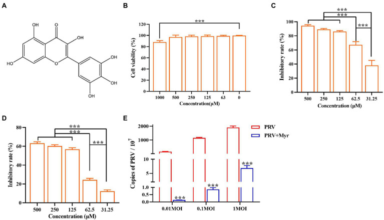
Anti-PRV activity of Myricetin. (A) The structural formula of myricetin. (B) The cell toxicity of myricetin. PK-15 cells were incubated with different concentrations of myricetin for 48 h, then the cell viability was measured by CCK-8. (C) The inhibitory rate of myricetin against PRV. PK-15 cells were infected with PRV (100TCID50) and treated with myricetin at the same time. After adsorption for 1 h, the mixture was replaced with fresh medium containing myricetin to culture for 48 h. Then a CCK-8 assay was performed. (D) The inhibitory rate of myricetin on PRV replication. PK-15 cells were infected with PRV (100TCID50) and treated with myricetin after 1 h post infection. After incubation for 48 h, a CCK-8 assay was used for detection. (E) MOI assay. PK-15 cells were infected with PRV (MOI = 0.01, 0.1, and 1, respectively) in the presence of myricetin; after incubated for 1 h at 37°C, the mixture was replaced with fresh compound-containing medium. The DNA copy of PRV was evaluated by FQ-PCR. Values are presented as means ± SD (n = 6). PRV, the infected group without treatment; PRV + Myr, the infected group with myricetin treatment. Symbol “***” represents p < 0.001 between the PRV group and PRV + Myr group.

