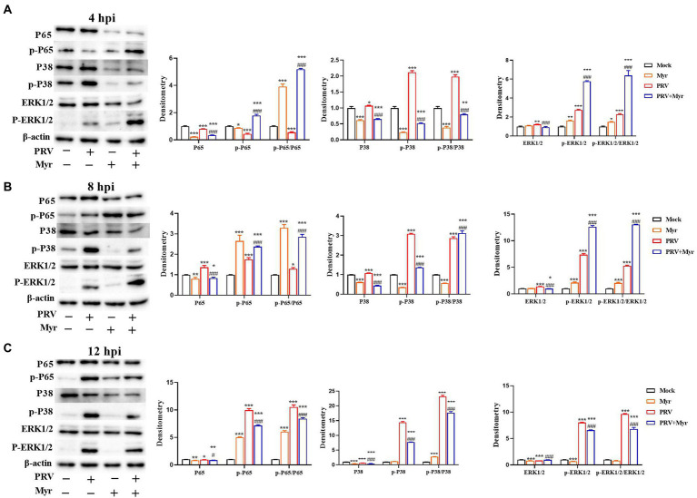
Figure 5.
The changes of the NF-κB and MAPK pathways. The PK-15 cells were infected with or without PRV (MOI = 1) in the presence or absence of myricetin. Expressions of P65, p-P65, ERK1/2, p-ERK1/2, P38, and p-P38 were determined by Western blotting at 4 (A), 8 (B), and 12 (C) hpi. Mock, the uninfected-untreated cells; Myr, the uninfected group with myricetin treatment. PRV, the infected group without treatment; PRV + Myr, the infected group with myricetin treatment. Data are presented as mean ± SD, n = 6. Symbols “*, **, and ***” represent p < 0.05, p < 0.01, and p < 0.001, respectively, between the mock group and other groups. Symbols “# and ###” represent p < 0.05 and p < 0.001, respectively, between the PRV group and PRV + Myr group.

