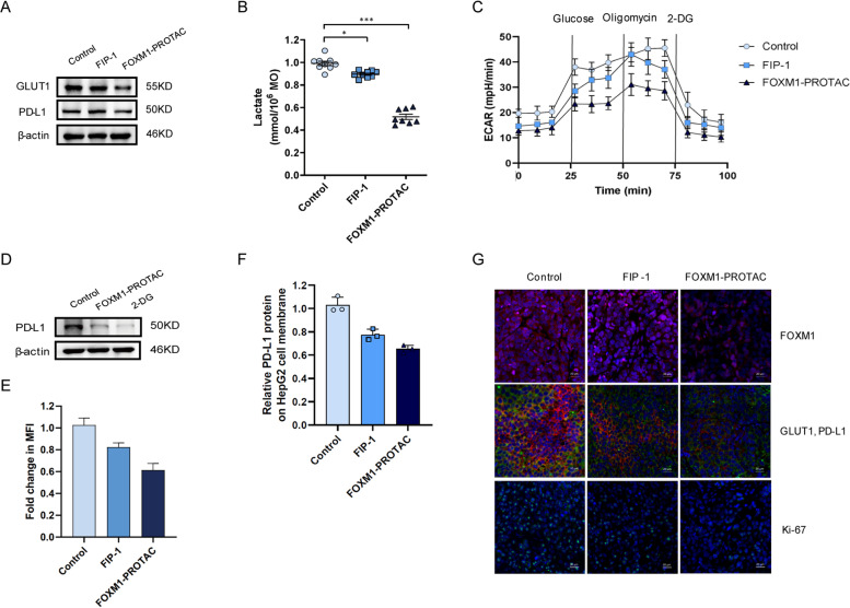
Fig. 6.
FOXM1-PROTAC inhibits the expression of GLUT1 and PD-L1. A Detection of PD-L1, GLUT1 in HepG2 cells treated with FIP-1 and FOXM1-PROTAC for 24 h. B Decreasing lactate level in HepG2 cells, treated with FIP-1 and FOXM1-PROTAC for 24 h, was showed by Microplate Reader. C, Seahorse extracellular flux analyzer (SEFA) measurement of ECAR metabolic profile in HepG2 cells treated with FIP-1 and FOXM1-PROTAC for 24 h. D Expression of PD-L1 in HepG2 cells after treating with 2-DG for 24 h. E HepG2 cells treated with FIP-1 and FOXM1-PROTAC for 24 h uptake analysis of 2-NBDG (200 μM) on a Flow cytometer. F Flow cytometer analysis of PD-L1 expression on tumor cell surface before or after FOXM1-PROTACs treatment. G Immunofluorescence of tumor samples, FOXM1 (red single), GLUT1 and PD-L1 (red and green merged), ki-67 (green single) and Cell nuclei was stained (blue)

