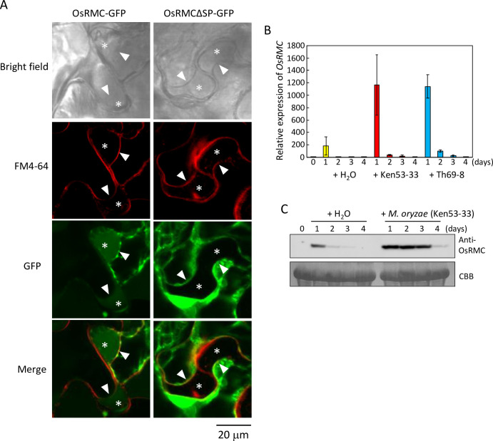
Fig 3. OsRMC is localized to apoplast and the gene expression is induced by M. oryzae infection.
(A) OsRMC is localized to apoplast. Subcellular localization of OsRMC was determined using plasmolyzed leaves of N. benthamiana overexpressing OsRMC-GFP or OsRMCΔSP-GFP by confocal laser scanning microscopy. The cells were also stained with FM4-64 to label the plasma membrane. Asterisk indicates the generated apoplastic space in the plasmolyzed cells. Arrow head indicates the position of plasma membrane. Bars, 20 μm. (B) OsRMC expression induced by M. oryzae infection in both compatible and incompatible interactions. Expression levels of OsRMC transcripts in rice (‘Hitomebore’) leaves after inoculation with M. oryzae (Ken53-33, compatible strain; TH69-8, incompatible strain, 3.0 X 10⁵ conidia each) were determined by qRT-PCR. OsRMC expression level was normalized using that of the rice ubiquitin gene (LOC_Os03g03920.1). Data are means ± SD of three independent determinations. (C) OsRMC protein expression is induced after M. oryzae infection. Protein extracts (20 mg) from rice leaves 0–4 days after M. oryzae (Ken53-33) inoculation were subjected to immunoblot analysis using an anti-OsRMC antibody. Equal protein loading on SDS-PAGE was verified by Coomassie Brilliant Blue (CBB) staining.

