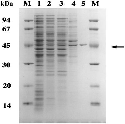
FIG. 1.
Purification of CitA from S. coelicolor crude extracts. Representative samples from each chromatography step (Table 1) were analyzed by SDS–12.5% PAGE. S. coelicolor crude extract (lane 1) was fractionated by DEAE-Sepharose (lane 2), Q-Sepharose (lane 3), and MonoQ anion-exchange chromatography (lane 4) and Matrex gel red A affinity chromatography to yield a pure preparation of CitA (lane 5; arrow). The molecular masses of the protein marker depicted in lane M are shown on the left.

