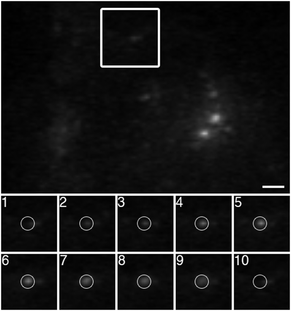
Figure 5.

Visualizing the arrival of VSVG-GFP to the PM. VSVG-GFP plasmid DNA was transported into neurons by electroporation prior to plating. At 7 days in vitro, the trafficking of VSVG-GFP vesicles to the PM was imaged by TIRFM using a 60× 1.45 NA objective at 11.32 frames per sec. The panels labeled 1 through 10 are sequential frames. As a VSVG-GFP vesicle nears the PM, it becomes brighter. Upon fusing with the PM, the signal rapidly diffuses. Bar = 2 μm.
