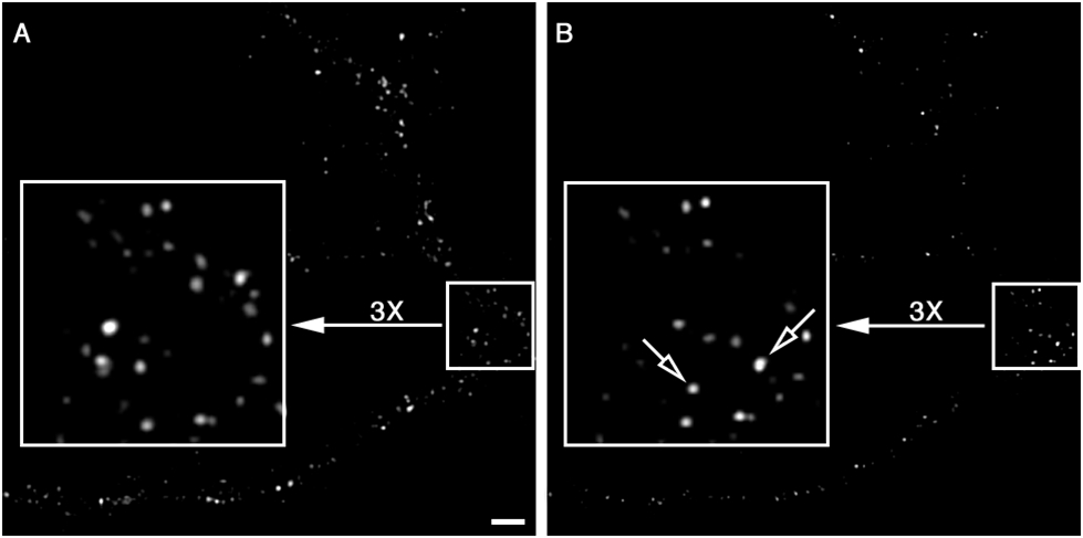
Figure 6.

Visualization of Y1r arrival at the PM by TIRFM. (A) Taken shortly after NPY-488 was added to the culture medium. The signal-to-background ratio is not affected by the presence of free fluorescently labeled agonist in the medium. (B) Captured 10 min after the image in A. Note the loss of 488 fluorescence along the dendritic branches and in some cases (open arrows) the detection of new receptors at the PM. Bar = 2 μm.
