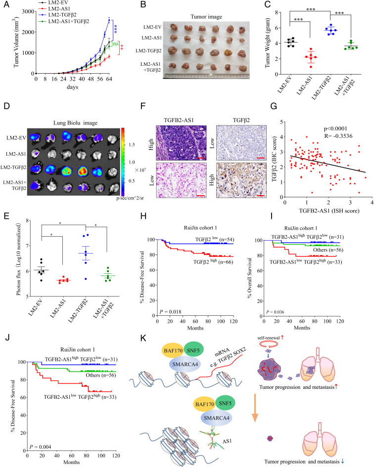
Fig. 8.
TGFB2-AS1 suppresses TNBC progression and metastasis via inhibiting TGFβ2. After tumor cells were injected into the mammary fat pad orthotopically, tumor growth curves were measured. (A) Tumors were harvested, photographed (B), and weighed (C) on day 64. IVIS images of lung metastasis (D) and luciferase quantitative data (E) on 64 d after orthotopic injection (n = 6 per group). Data are presented as means ± SEM (n = 6). Statistical significance was assessed using two-tailed Student’s t test. (F) Representative images (scale bar, 50 μm) of ISH of TGFB2-AS1 and immunohistochemical staining of TGFβ2 in Ruijin cohort 1. (G) Correlation between TGFB2-AS1 and TGFβ2 expression from RuiJin cohort 1 determined by Pearson correlation analysis. (H) Kaplan–Meier analysis of disease-free survival of TGFB2 from Ruijin cohort 1. (I and J) Kaplan–Meier analysis of the overall survival and disease-free survival of TGFB2-AS1high/TGFβ2low and TGFB2-AS1low/TGFβ2high from Ruijin cohort 1. (K) Schematic view of the regulatory mechanisms and roles of TGFB2-AS1 in suppressing TNBC progression. Error bars represent mean ± SEM. Statistical significance was assessed using two-tailed Student’s t test. *P < 0.05. **P < 0.01. ***P < 0.001. ns, not significant.

