Fig. 5.
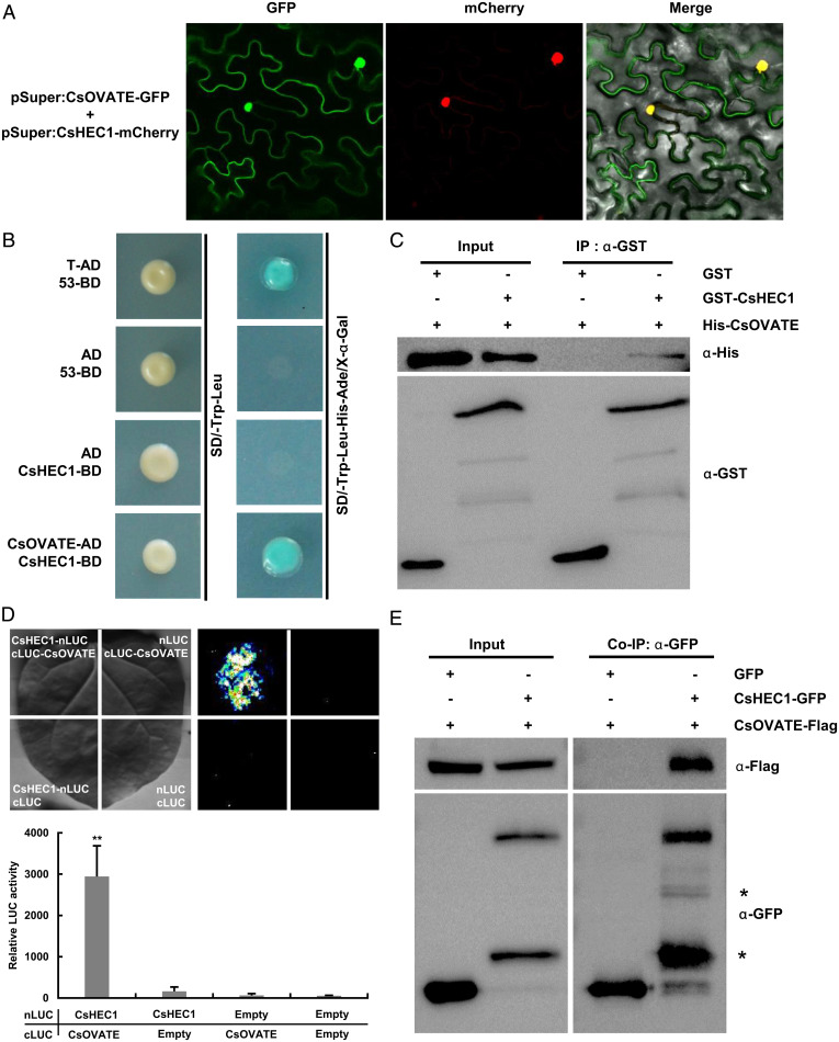
CsHEC1 directly interacts with CsOVATE at the protein level. (A) CsOVATE colocalization with the CsHEC1 protein in N. benthamiana leaves. The yellow signal in the merged field represents the colocalization signal in the nucleus. (B) Interaction of CsHEC1 and CsOVATE in the yeast two-hybrid system. The combination of T-AD and 53-BD was used as a positive control. (C) CsHEC1 interacts with CsOVATE in vitro tested by a GST pull-down assay. The combination of GST and His-CsOVATE was used as a control. (D) Firefly luciferase complementation imaging analysis. CsHEC1-nLUC and cLUC-CsOVATE were transiently coexpressed in N. benthamiana, and the remaining combinations were used as controls. Representative pictures (Top) and the relative luciferase activity value are shown (Bottom). Values are means ± SD (n = 6). Two-tailed Student’s t test (**P < 0.01). (E) Co-IP assay showing that CsHEC1 interacted with CsOVATE in vivo. The total and precipitated proteins were detected by immunoblotting using anti-GFP antibody or anti-Flag antibody. The asterisks indicate nonspecific bands.

