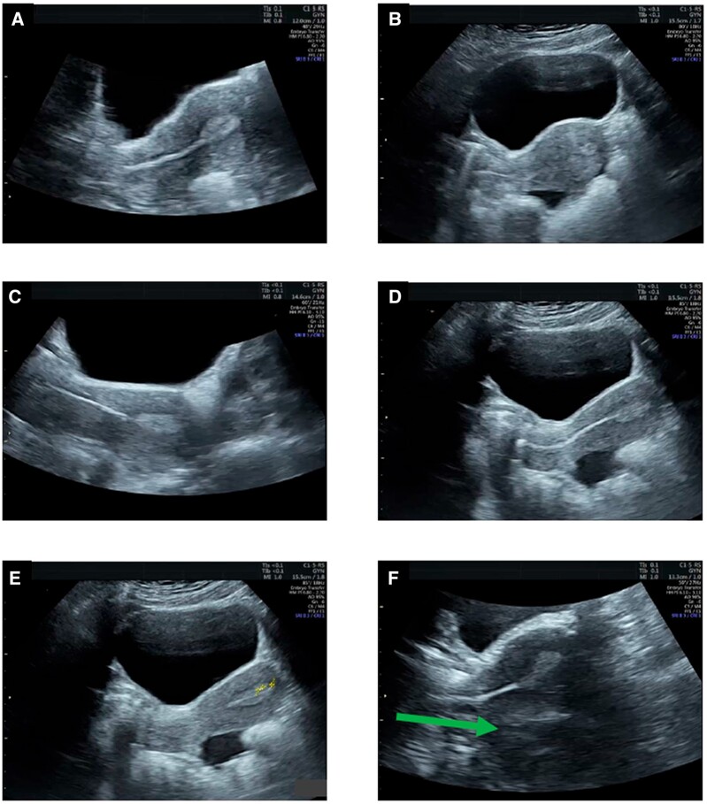
Figure 8.
Ultrasound images showing various factors related to a transabdominal ultrasound-guided embryo transfer. (A) Zoom window focused on the uterine cavity. (B) Deep field view of embryo transfer (ET) during catheterization. (C) Retroverted uterus ET. (D) ET catheter approaching the fundal part of the endometrium. (E) Measurement of distances between tip of catheter and fundal endometrium and between embryo bubble and endometrium. (F) Artefacts during ET: example of mirror image artefact (uterus is anteverted but there is a false image showing a retroverted uterus).

