Fig. 2.
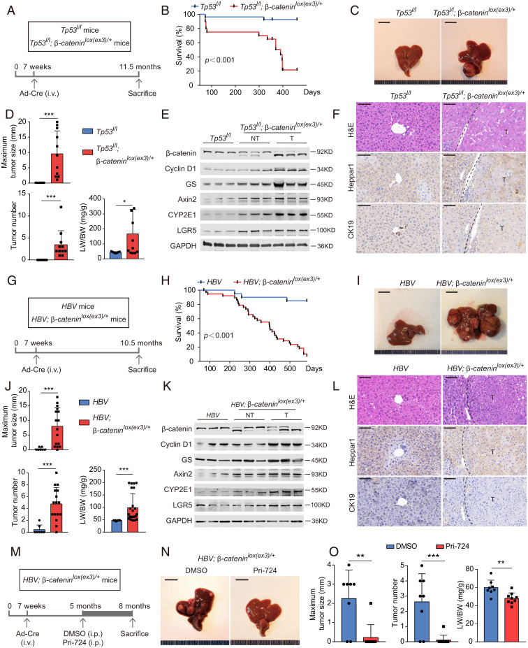
Oncogenic β-catenin colludes with distinct carcinogenic factors in furtherance of hepatocarcinogenesis. (A–F) β-Catenin activation and Tp53 deletion in mice. (A) Tp53l/l and Tp53l/l; β-cateninlox(ex3)/+ mice were injected with Cre-adenovirus via tail vein 7 wk after birth. (B) Survival of Tp53l/l mice (n = 27) and Tp53l/l; β-cateninlox(ex3)/+ mice (n = 20). (C–F) Representative pictures (C), the maximum liver tumor size (Upper), tumor number (Lower Left), ratio of liver weight to body weight (Lower Right) (D), immunoblotting (E), and representative H&E, Heppar1 and CK19 stainings (F) of liver tissues from 11.5-mo-old mice. (G–L) Mutant β-catenin and transgenic HBV in mice. (G) HBV and HBV; β-cateninlox(ex3)/+ mice were injected with Cre-adenovirus to generate HBV and HBV plus hepatic β-catenin mutant mice. (H) Survival of HBV mice (n = 21) and HBV; β-cateninlox(ex3)/+ mice (n = 39). (I–L) Representative pictures (I), the maximum liver tumor size (Upper), tumor number (Lower Left), ratio of liver weight to body weight (Lower Right) (J), immunoblotting (K), and representative H&E, Heppar1 and CK19 stainings (L) of liver tissues from 10.5-mo-old mice. (M–O) Pri-724 treatment. (M) HBV; β-cateninlox(ex3)/+ mice were injected with Cre-adenovirus by 7 wk, treated with vehicle (DMSO, dimethyl sulfoxide) or Pri-724 by 5 mo and killed by 8 mo after birth. (N) Representative liver pictures of 8-mo-old mice. (O) The maximum liver tumor size (Left), tumor number (Middle), and ratio of liver weight to body weight (Right) were analyzed. Data are shown as mean ± SD *P < 0.05, **P < 0.01, ***P < 0.001.

