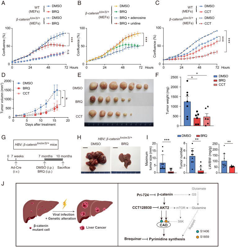
Fig. 7.
Suppression of AKT2-activated pyrimidine synthesis abrogates oncogenic β-catenin–mediated cell proliferation and tumorigenesis. (A–C) Cell proliferations of wild-type and β-cateninΔ(ex3)/+ MEFs were measured with Incucyte assay. (D–F) Nude mice with subcutaneously inoculated β-cateninΔ(ex3)/+ MEFs were treated with DMSO, BRQ (25 mg/kg, three times/wk), or CCT (25 mg/kg, five times/wk). (D) Tumor growth was plotted as the mean change in tumor volume. Tumor graphs (E) and tumor weights (F) at the end of treatment. (G) The 7-wk-old HBV; β-cateninlox(ex3)/+ mice were injected via tail vein with Cre-adenoviruses. DMSO or BRQ treatment was started when mice were 7-mo-old and killed 3 mo later. (H) Representative livers from 10-mo-old HBV; β-cateninlox(ex3)/+ mice. (I) The maximum liver tumor size (Left), tumor number (Middle), and ratio of liver weight to body weight (Right) were analyzed. (J) Schematic illustration of oncogenic β-catenin stimulating pyrimidine synthesis in promotion of hepatocarcinogenesis. *, #P < 0.05. Analysis was performed using t test (A–D, and I) and one-way ANOVA (F). Data are shown as mean ± SD.

