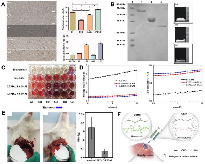
Figure 5.
(A) Platelet adhesion and fibrinogen concentration in response to silk fibroin and SF-PEG sponges. Image adapted from Wei et al. [134]. (B) SDS-PAGE showing three hemostatic peptides and their sponges after purification. Image adapted from Yang et al. [136]. (C) PDA–sodium alginate–polyacrylamide (PDA–SA–PAM) hydrogel network showing accelerated clotting time [144]. (D) The addition of polyDOPA to SA–PAM structure improves rheological properties drastically. Images adapted from Suneetha et al. [144]. (E) Hemostatic capacity of the triblock DOPA peptide. Image adapted from Lu et al. [142]. (F) Hemostatic OCMC and antimicrobial G3KP polysaccharide-peptide dendrimers for rapid curing tissue adhesive-hemostat development. Image adapted from Zhu et al. [147].

