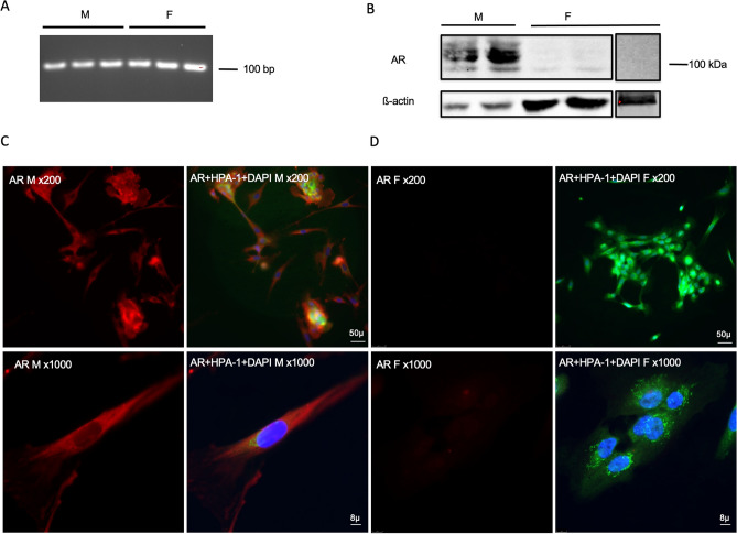
Figure 3.
Identification of AR in human conjunctival goblet cells cultured from males and females. RNA was isolated from cultured goblet cells and RT-PCR performed using primers for human Ar. A single band was detected at around 100 bp in samples from both males (M) and females (F) (A). Protein samples were collected from cell pellets and Western Blot analysis was performed using antibody against AR. A major band at 100 kDa was detected in males while no immunoreactivity was detected in females (B). Immunofluorescence microscopy was performed on cultured goblet cells using antibodies to AR (C and D). (C) Indicate immunofluorescence to AR (red) in male cells; (D) indicate immunofluorescence to AR (red) in female cells; an overlay of anti-AR, HPA-1 (green, indicates the secretory granules of goblet cells), and DAPI (blue, indicates cell nuclei) is shown next to the single channel image of AR. Magnification was × 200 in the upper panels; × 1000 in the lower panels. Original blots/gels are presented in Supplementary Figs. 5 and 6.

